INTRODUCTION
Bilateral vocal cord palsy is one of the causes that trigger nocturnal stridor and obstructive sleep apnea (OSA), and three cases of OSA due to bilateral vocal cord paralysis have been described in the medical literature. In 1987, Ruff et al.1 reported a case of bilateral vocal cord palsy and sleep apnea in a patient with type I Chiari malformation. In 1996, McBrien et al.2 reported a case of bilateral vocal cord palsy in a patients with Shy-Drager syndrome (multiple system atrophy with autonomic phenomena) who sought treatment for nocturnal stridor and sleep apnea. In 2003, Luaazy and Hasse described a patient with breathing difficulty, snoring and severe OSA which were caused by bilateral vocal cord palsy of unknown origin.3 In addition to vocal cord palsy, there were several reports that congenital and acquired factors have resulted in a collapse of glottis structures that, in turn, has resulted in laryngeal stridor and OSA.4,5 In this aspect, the role of laryngeal structure, including vocal cord, to develop laryngeal stridor and OSA is important. However, the role was overlooked by many clinicians. And laryngeal exam was not routinely performed to investigate the cause of OSA. In this article, we describe a case of nocturnal stridor with combined OSA which was caused by undiagnosed bilateral vocal cord palsy and review the importance of laryngeal exam to find the problems of glottis structures.
CASE
A 65-year-old man was referred to the ENT department for evaluation of 20-year history of snoring, witnessed apneas, excessive daytime sleepiness. He visited several local hospitals for his problems. However, his symptom did not get better. Four days ago, he visited emergency department due to dysarthria and right side motor weakness. He was diagnosed with pure motor stroke and has been hospitalized to the neurology department. He had 10-year history of hypertension and type II diabetes, but he has only taken medication for hypertension. Five years ago, he had a history of left side motor weakness and dysarthria. At that time, he sought herbal medication and has fully recovered. One year ago, his breathing difficulty was aggravated when he ascended the stairs, and visited a local hospital and diagnosed with asthma. His breathing difficulty was controlled with topical steroid therapy. However, he complained with dyspnea on exertion and voice change when he exercised hard.
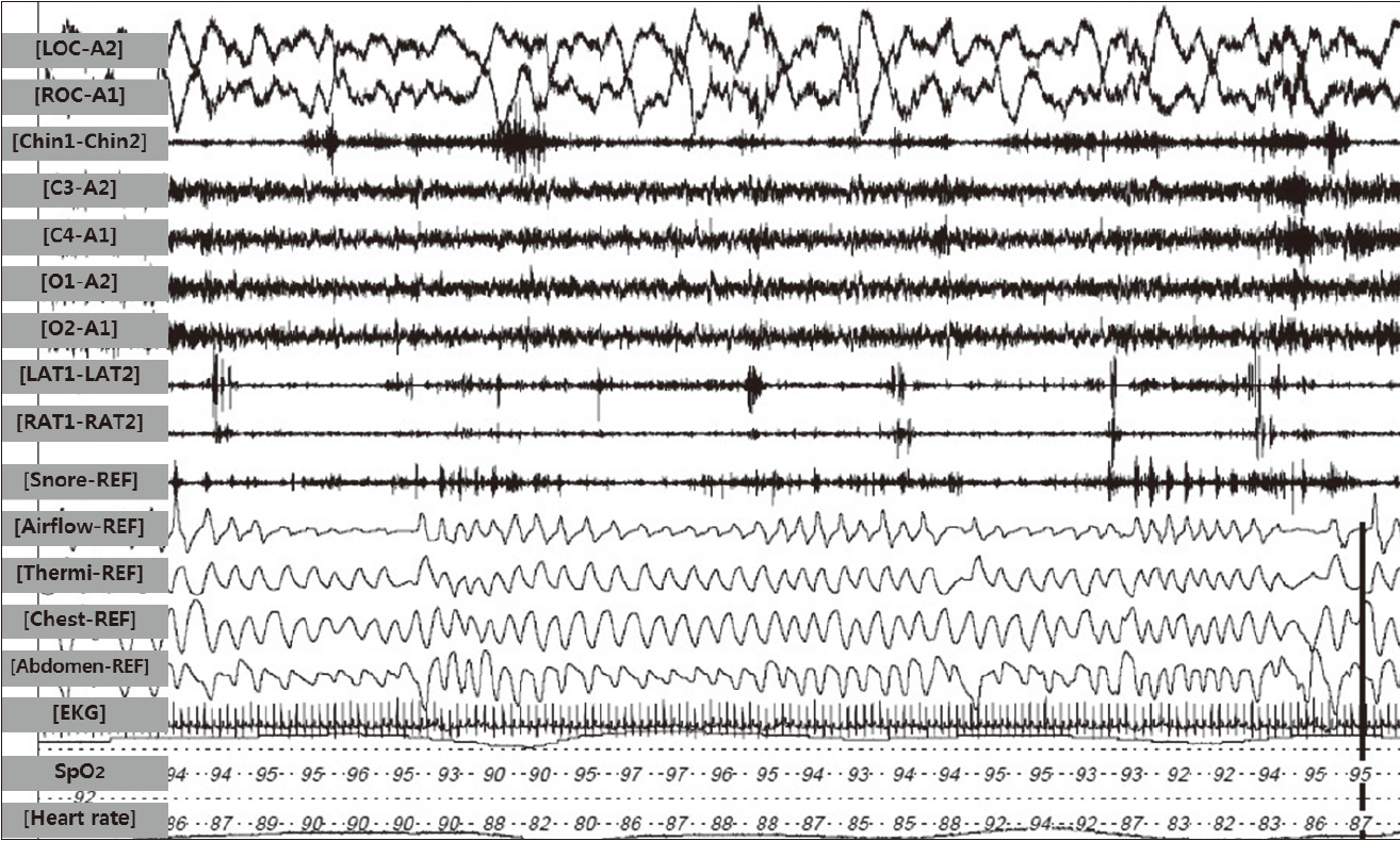
When we met him, he has fully recovered from motor weakness and dysarthria after 3 days hospital care. He exhibited no signs of any peripheral or central nervous system disease, and other findings were normal. Nocturnal polysomnography over full night revealed that his apnea/hypopnea index was 25 and his minimum arterial oxygen saturation (SaO2) level was 88% (Fig. 1). Mean SaO2 was normal during wakefulness (95%) and only slightly reduced during sleep (88%). In addition, harsh high-pitched nocturnal breathing noise was also reported. Pulmonary function test was performed on him twice, but the test failed due to his breathing difficulty. The physical examination showed that the patient’s weight was 72 kg, height was 164 cm [body mass index = 26.8 kg/m2], and his neck circumference was 42 cm. He had a little bit macroglossia, but didn’t have enlarged tonsils or other anatomic abnormalities on his oral cavity. Nasopharygeal examination and Müller maneuver using flexible fiberoptic laryngoscopy didn’t reveal any abnormalities. On examination of glottis structure, we discovered that he had bilateral vocal cord palsy, which showed the paramedian fixation. We evaluated the cause of his vocal cord palsy using computed tomographic (CT) angiography on head and neck, brain MRI, chest simple x-ray, neck CT, but we couldn’t find that. So he was diagnosed with nocturnal stridor with combined moderate OSA caused by bilateral vocal cord palsy of unknown origin. He underwent tracheostomy under local anesthesia. His condition was improved considerably after the operation, and for the first time, since the finding of the symptom, he was able to sleep without snoring and sleep apnea. During the follow-up, the quality of the sleep was better and his vocal cord movement has progressively improved. After he had successful rehabilitation for vocal cord palsy about one and half year, his tracheostomy cannula removed. His snoring, sleep apnea and breathing difficulties were completely disappeared.
DISCUSSION
There are several well-known anatomic abnormalities that are responsible for OSA. These are adenotonsillar enlargement, macroglossia, low position of the hyoid bone or excessive length of the soft palate.6 The role of the larynx in the above-mentioned field has not been fully determined. The mechanism of snoring in patients with bilateral vocal cord paralysis seems to be similar to OSA syndrome. The difference consists in the level of the flow limitation. In order to push the obstruction in the glottis, the patient has to create a higher inspiratory pressure and hence the dynamic compression that makes inspiration more difficult increases.
There were some cases that congenital or acquired factors have resulted in a collapse or dysfunction of laryngeal structures, which, in turn, has resulted in the development of sleep-related stridor and OSA. As previously mentioned, Ruff et al.,1 McBrien et al.2 and Luaazy and Hasse3 each reported a case of sleep-related stridor and OSA that was caused by bilateral vocal cord paralysis. In addition, Woo reported two cases of progressive OSA in which the epiglottis prolapsed after surgery on the larynx and pharynx,4 and Bonilla et al.5 reported a case of OSA in a child with epiglottic aplasia. Sameer et al. demonstrated the improvement of sleep apnea and growth retardation after a surgical correction of children who had OSA caused by bilateral vocal cord palsy and laryngomalacia.7 It is well known that any space-occupying lesions between pharynx and glottis can cause obstruction and predispose a patient to laryngeal stridor and OSA.8
In the evaluation of OSA patients, the physical examination of mouth, nose, and throat for extra or large tissue was performed routinely and easily at the sleep clinics. However, laryngeal examination was not done usually and many physicians overlooked the larynx problems that cause OSA. It is because not only the laryngeal examination is not so easy but also laryngeal problems to cause OSA are rare. In this case, the patient may be treated with continuous positive airway pressure depending on the results of his sleep study, nose and pharynx examination. If we don’t perform the laryngeal examination, we couldn’t find the cause of nocturnal stridor with combined OSA and bilateral vocal cord palsy. In addition, his daytime symptoms usually were episodic during daytime, but could be aggravated and presented constantly showing various symptoms. However, the follow-up polysomnography was not performed, so we couldn’t get the objective data after the improvement of vocal cord palsy and decanulation. This is the limitation of this study. Through this case, we emphasize that laryngeal examination was so important and should be performed routinely to evaluate the cause of sleep-related stridor and OSA.